Orthopedics & Osteopathy
in the Algarve, Portugal
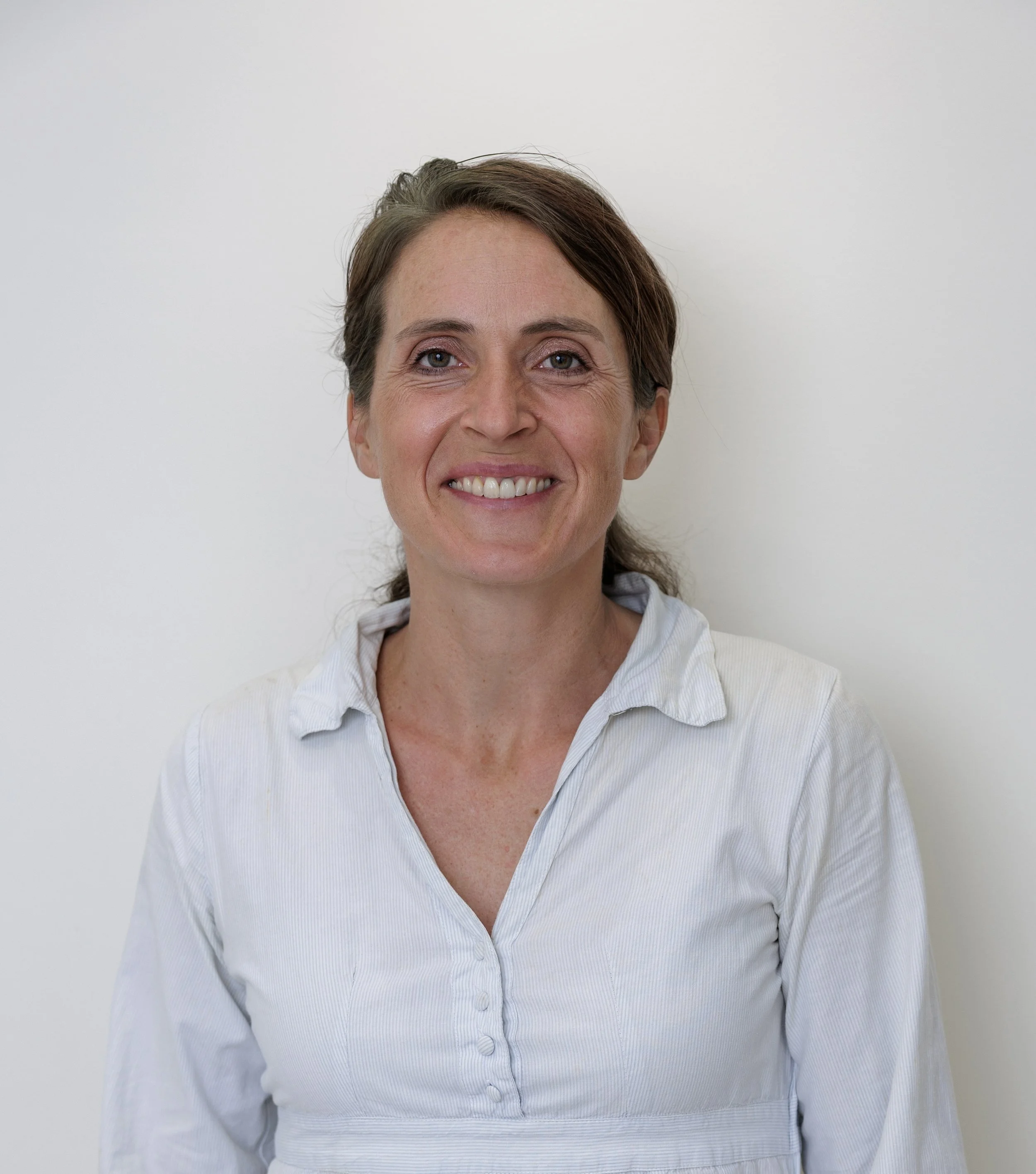
“I am offering a holistic approach to orthopedic medicine with a focus on a thorough diagnosis and different treatment options from modern orthopedic medicine to chirotherapy and osteopathy”
About me
-
2023- present
Private Practice, Family Medical Centre & Antiaging Clinic, Lagos- Portugal
2021- present
Private Practice, Clinica Medin, Lagoa- Portugal
2019- present
Private Practice, Family Medical Centre & Antiaging Clinic , Quinta do Lago- Portugal
2012- present
Klinik Manhagen, Hamburg- Germany
2017- 2025
Private Practice, German Medical Centre, Carvoeiro- Portugal
2017
Specialist Exam in Orthopedic and Trauma Surgery
2015- 2017
Private Practice, Orthopädiezentrum Kurfürstendamm, Berlin- Germany
2014
Further Education in osteopathic medicine with certificate „doctor of osteopathy“
DO of the german/american organisation DAAO
2012- 2013
Department of Shoulder Surgery, Sports- Trauma- and Arthroscopic Surgery,
Roland Klinik Bremen- Germany
2010- 2012
Department of Orthopedic and Trauma Surgery, Asklepios Klinik Nord Heidberg- Germany
2009- 2010
Department of Anesthesia and Intensive Care, Sana Klinikum Sommerfeld- Germany
2009
PhD at University Hamburg- Germany
2008- 2009
Department of Arthroplasty, Sana Klinikum Sommerfeld- Germany
2002- 2008
Medical school at University of Hamburg and Tauranga, New Zealand
-
Specialist in Orthopedics and Trauma Surgery
Doctor of osteopathy (DAAO)
Certified FDM- Therapist (fascial distorsion model of Typaldos)
Manual Medicine MWE
Foot Surgery GFFC
Emergency Medicine
ATLS Instructor (advanced trauma life support)
Certified 800hrs yoga teacher
-
German
English
Portuguese


Direct booking with the clinic
-
-
R. José Ferreira Canelas 40
8600-744 Lagos
-


Contact me directly

Specialty
Treatment of Osteoarthritis
Hyaluronic Acid vs. Platelet-Rich Plasma vs. Orthokine®
Osteoarthritis, or joint degeneration, is one of the most common diagnoses in orthopaedic practice. The knee, hip and joints of the hands are particularly susceptible to wear and degeneration, but any joint can be affected, causing pain and restricted movement and thereby reducing a patient’s quality of life.
Typical symptoms of osteoarthritis include pain when starting to move and during weight-bearing activities, particularly in the morning and after longer periods of rest, often accompanied by stiffness. Pain and stiffness usually improve after the first movements in the morning but may return or increase with prolonged activity. In more advanced stages, patients may also experience pain at rest and during the night.
The typical restriction in movement results from the loss of cartilage within the joint as well as changes in the joint capsule, tendons and muscles surrounding the joint.
Diagnosing osteoarthritis requires a careful medical history, a physical examination and, if necessary, an X-ray.
There are various treatment options available. Maintaining a healthy body weight and avoiding excessive or inappropriate strain on the affected joint can often help to reduce pain.
Activities that place less stress on the affected joint, such as cycling or swimming, are recommended. Walking on softer surfaces, such as forest paths or grass, possibly with the use of walking poles or crutches, may also provide relief.
Physical measures such as ice or cold applications for acute pain or swelling, as well as heat applied to the surrounding muscles, can be soothing and help relieve symptoms.
Physiotherapy and, for example, acupuncture are important complementary treatments. Acute pain and significant inflammation during an arthritic flare-up can temporarily be treated with non-steroidal anti-inflammatory drugs (NSAIDs), provided there are no contraindications, such as certain cardiovascular or gastrointestinal conditions or blood-clotting disorders.
If these measures do not provide sufficient relief, injection therapies into the affected joint may be considered.
There are several different injection treatment options.
Treatment with Platelet-Rich Plasma (PRP) has shown good results in patients with mild to moderate osteoarthritis. For this treatment, blood is drawn from a vein in the arm. Through centrifugation and the use of an anticoagulant, the plasma is separated from the other blood components. The plasma, which contains anti-inflammatory components and growth factors that support regenerative processes, is then injected into the joint under sterile conditions, where it can act on the cartilage and the often inflamed synovial membrane.
PRP therapy can also be very effective in the treatment of overloaded or chronically inflamed tendons, such as tennis elbow, Achilles tendon irritation and muscle injuries.
In cases of moderate or advanced changes to the joint cartilage, the lubricating, shock-absorbing and nutritional functions of the synovial fluid can be improved with hyaluronic acid injections. Usually, a single injection is administered and may provide symptom relief for approximately 6–12 months.
In cases of advanced osteoarthritis, particularly when inflammation is a predominant factor and/or when hyaluronic acid injections have not provided satisfactory results, treatment with Orthokine® may be considered. This treatment is usually administered as 3–4 injections, approximately one week apart, into the affected area (see below).
Surgery involving the implantation of an artificial joint replacement should generally only be considered in cases of so-called treatment-resistant osteoarthritis, when conservative treatments have not provided sufficient relief and the patient has a strong desire to proceed with surgery.
Ultimately, the decision always lies with the patient, who should weigh the potential improvement in quality of life against the risks and expected outcome of surgery.


Specialty
Golfers and Tennis Elbow
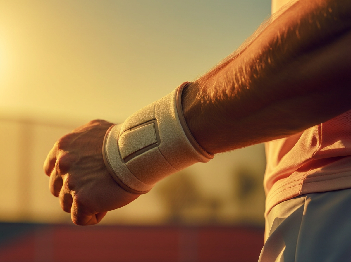
The frequent medical complaint of a tennis elbow with pain on the outer side of the elbow (called epicondylitis humeri radialis) and the slightly less common problem of a golfer’s elbow with pain on the inner part of the elbow (epicondylitis humeri ulnaris) are caused by painful tissue changes in the tendons of the elbow through repetitive movement of the wrist- and hand extensors and flexors.
Often this complaint is caused by playing tennis and golf but this phenomenon is also found in people working in maintenance, long hours on the computer keyboard, in cleaning jobs, athletes in ball sports, but it is also more generally caused by intense monotonous straining of the corresponding muscles of the forearm. An uncommon exposure (f.e. a house renovation) or a rapid increase of a particular strain (f.e. starting training after a break) are the cause of the complaints. This strain might lead to inflammation of the sinewy area at the transition of bones to muscle structure at the elbow.
Not only the shortening and the hypertension of the muscles of the forearm, also a missing stabilization and unfavorable coordination of the shoulder girdle or changes in the cervical spine caused by degeneration or blockages lead to the longer lasting symptoms of epicondylitis.
Pain caused by inflammatory processes and tiny injuries, so called micro-injuries, initially start while straining the arm and later on will also occur in resting mode. Typically the pain radiates along the muscular tissue to the forearm.
A diagnosis can already be made with a thorough examination. In the examination and in the subsequent treatment it is important to include the shoulder girdle, the cervical spine and the nerve function.
The treatment should be holistic. Measures to ease the pain like local heat or cold, ointment with anti-inflammatory creams, f.e. with comfrey extract or nightly curd compresses are beneficial. Plant-based anti-inflammatory preparations to take internally, like bromelain or curcurmin and in persistent cases also the intake of traditional anti-inflammatory painkillers like Ibuprofen is commonly advised.
Accompanied by manual treatment of the tendon insertions and treatment of tensed muscles by a physiotherapist, preferably in combination with physical methods like ultrasound therapy.
Special exercises for stretching and relaxation are important, also in form of daily self practice (concentric muscle training). The increasingly popular fascia roll is able to support the relaxation of tensed muscles and will help to continue the work of the therapist.
Also, the so called epicondylitis-brace, worn close to the elbow on the forearm, diverts the pull of the muscles from the origin at the elbow to less strained areas and can provide relief.
Well applied kinesiotapes bring about an increased blood circulation of the affected area and increase a relaxation of the strained muscles underneath through permanent relocation of connective tissue.
Athletes should always have their techniques inspected by an experienced coach in order to correct postural deficits.
Osteopathy with local and holistic treatment is very helpful to shorten the duration of symptoms which often last several months.
A set of specific exercises to practice at home are often passed on to the patient.
Shots of cortisone and operations meanwhile are normally not recommended any more.
For athletes a slow progression of strain of any kind (so called pacing) and continuous stretching and relaxation of muscles proved very effective to prevent recurring injuries.
In long histories of complaints injections with platelet rich plasma extracted from the patients own blood show excellent results to speed up the healing.
Specialty
Shockwave Therapy – a modern treatment for tendon and muscle pain
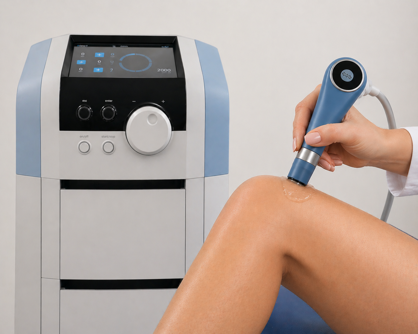
Radial shockwave therapy is a modern, non-invasive treatment used for tendon problems, such as calcifications or tendon irritation, as well as muscle tension.
It has been used successfully for many years in orthopedics, sports medicine, and physiotherapy.
How does radial shockwave therapy work?
During the treatment, high-energy pressure waves are applied to the affected area using a handheld device. These impulses stimulate blood flow, metabolism, and cell activity, helping the body heal naturally.
The therapy can:
• improve blood circulation
• reduce pain
• relax muscles
• activate metabolism
• reduce inflammation
Especially in chronic conditions, shockwave therapy can help loosen tight tissue or reduces the inflammation in tissues and support healing.
Advantages of shockwave therapy
A major advantage is that the treatment is non-invasive.
It is done on an outpatient basis and usually takes only a few minutes. Many patients feel noticeable improvement after just a few sessions. Typically, 6–8 treatments are recommended.
Common uses
Shockwave therapy can help with many orthopedic and muscle problems, for example:
• muscle tension
• trigger points
• heel spur and plantar fasciitis
• Achilles tendon problems
• tennis elbow
• shoulder pain and tendon calcification
• neck and back pain
• tendon irritation
• overuse injuries
• sports-related injuries
Specialty
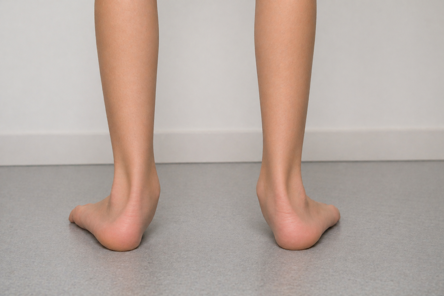
Toe Walking in Children
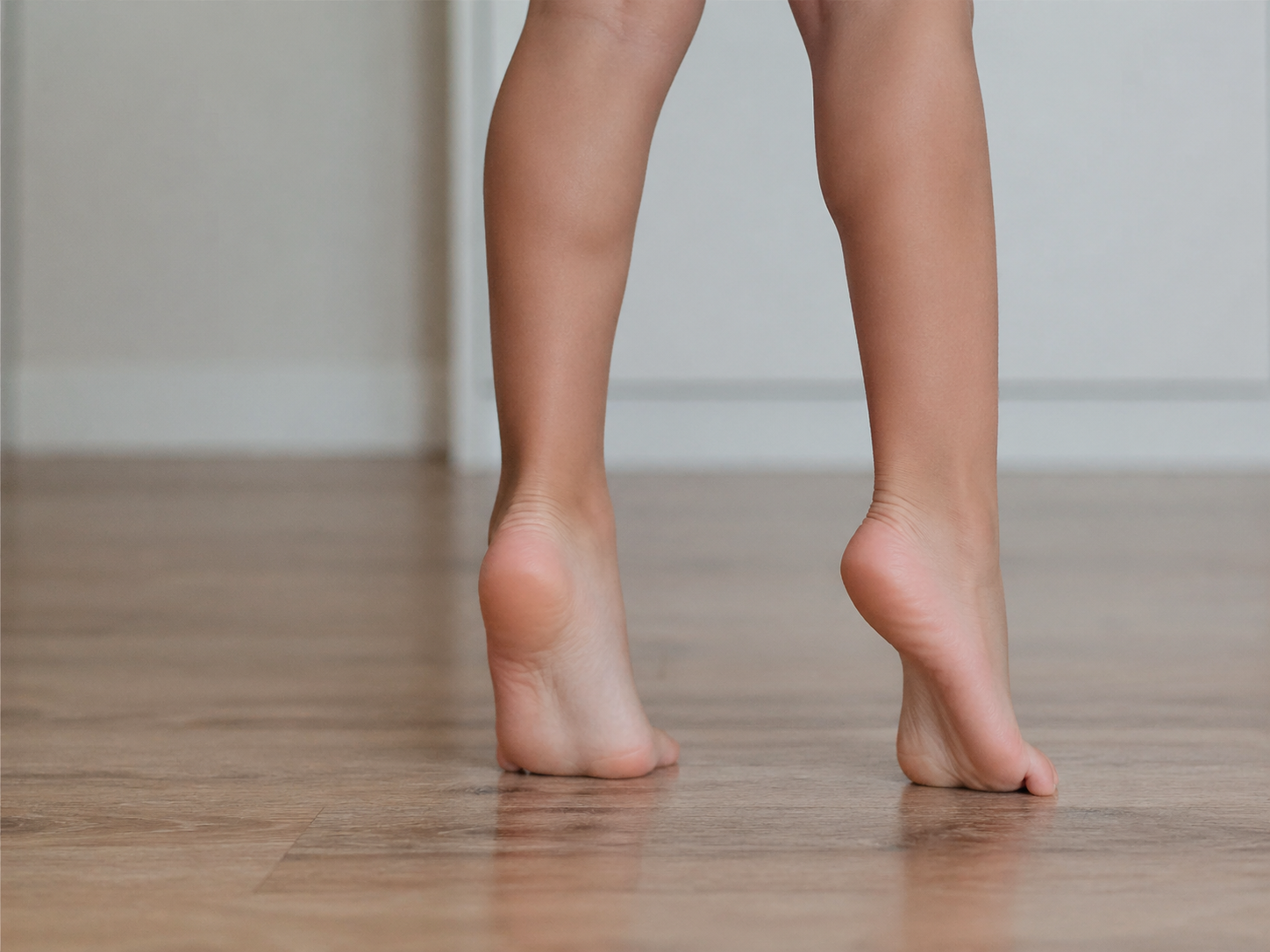
Many children walk on their toes at times.
In early childhood, this can be part of normal motor development. However, if toe walking continues over a longer period or becomes more noticeable as the child grows, a targeted medical evaluation is recommended.
For a long time, so-called “idiopathic toe walking” was considered simply a habit. More recent scientific findings – including the work of David Pomarino – suggest that neurological, neuromuscular, or genetic factors often play an important role in affected children.
Toe Walking Is Often a Symptom – Not a Diagnosis
Toe walking initially describes only a noticeable walking pattern: the child mainly walks on the front of the foot without fully placing the heel on the ground. There can be many different underlying causes.
From a clinical perspective, it is especially important to identify possible neurological or muscular connections early on. Children who walk on their toes often show signs of:
genetic predispositions
hereditary neuropathies
muscular developmental disorders
sensory processing differences
neuromuscular conditions
or central coordination difficulties
Conditions That May Be Associated with Toe Walking
Persistent toe walking may occur in connection with:
hereditary motor and sensory neuropathies (e.g. Charcot-Marie-Tooth disease)
mild forms of cerebral movement disorders
hereditary muscle diseases and myopathies
abnormalities in muscle tone
autism-related movement patterns
sensory integration difficulties
Even when children initially appear typical, closer examination often reveals subtle differences in muscle tone, coordination, balance, or motor control.
Long-standing toe walking can affect overall physical development. Over time, this may lead to:
shortening of the calf muscles and Achilles tendon
reduced ankle mobility
changes in foot structure
postural differences
knee, hip, or back pain
balance and walking difficulties
The earlier underlying causes are identified, the more targeted treatment can be. Modern assessment therefore includes not only gait observation but also neurological, orthopedic, and functional evaluation.
In children with toe walking, we perform a comprehensive clinical assessment, including:
movement patterns and gait analysis
muscle tone and strength
joint mobility
coordination and balance
neurological signs
family history
indications of neuromuscular conditions
If necessary, we recommend additional neurological or genetic testing to identify possible causes early.
Treatment is always based on the underlying cause and the child’s individual development. The goal is not only to change the walking pattern but to improve overall motor function.
Depending on the findings, treatment may include:
night splints and specific insoles
specialized physiotherapy
neurological follow-up
interdisciplinary developmental assessment
minimal invasive surgery
Early and well-differentiated evaluation can help prevent functional limitations and support healthy motor development in the long term.


Specialty
When the back is in pain
The Upper and Lower Cross Syndrome
Neck and back pain are increasing significantly in our society today. Almost everyone has experienced neck pain or lower back pain at some point in their lives. Not only prolonged sitting, computer work, poor posture, and lack of exercise contribute to these issues, but stress is also a common cause of these complaints. A skilled therapist or practitioner can identify recurring muscular imbalances, known as dysfunctions, by observing the patient's standing and sitting posture and performing a few palpation techniques.
In cases of neck pain, the therapist typically observes a rounded back, with the head and shoulders slightly forward and the neck compensating with increased rounding. This is called Upper Cross Syndrome or upper crossed syndrome. In this condition, the muscles in the front of the neck and between the shoulder blades (which retract the shoulder blades) are weak, while the trapezius muscle (also called the hood muscle) and chest muscles are shortened, tense, and tight. This can lead to various complaints, including neck pain, numb fingers, radiating shoulder pain, migraines, dizziness, tinnitus, or a bothersome sensation of tension.
Lower Cross Syndrome is characterized by weak abdominal and gluteal muscles and shortened and tense back and hip flexor muscles. This imbalance is particularly common in individuals who sit for long periods with minimal movement and can cause back pain, radiating leg pain, muscular tension, stiffness, and sometimes even abdominal complaints due to the tense hip flexor (also known as the psoas muscle).
Relief can be achieved through muscle relaxation performed by a physiotherapist or osteopath, the application of kinesiology tape on the tense muscles, or, in severe cases, injections of local anesthetics into highly tense muscles. Regular exercises with the increasingly popular fascia rollers also aid in relaxing tense muscles. However, regular stretching of the shortened and tense muscles and strengthening of weak muscles is essential. Improving posture by standing and sitting upright and simultaneously activating the muscles between the shoulder blades through retraction and pushing the head back from the forward posture to the neutral position above the spine can alleviate Upper Cross Syndrome symptoms. Additionally, it is important to stretch tense muscle groups, such as the trapezius or chest muscles.
For Lower Cross Syndrome, activating the abdominal muscles and training the rotation of the upper body using the core muscles for stabilization helps. Stretching and thus relaxing the tense hip flexor, while simultaneously strengthening the weak gluteal muscles, are crucial components of the training therapy. The best results can be achieved by consciously adjusting posture and regularly training with a qualified therapist who can guide the exercises from the beginning and correct any small errors in execution. However, even regular brisk walking with an upright posture and using the arms during walking or regular swimming can lead to initial muscle activation. It is advisable to rest only during acute pain conditions and preferably for a short period. Because it is in movement that the first successes are seen.