Orthopedic & Osteopathy
Consultations
in the Algarve, Portugal
“I am offering a holistic approach to orthopedic medicine with a focus on a thorough diagnosis and different treatment options from modern orthopedic medicine to chirotherapy and osteopathy”
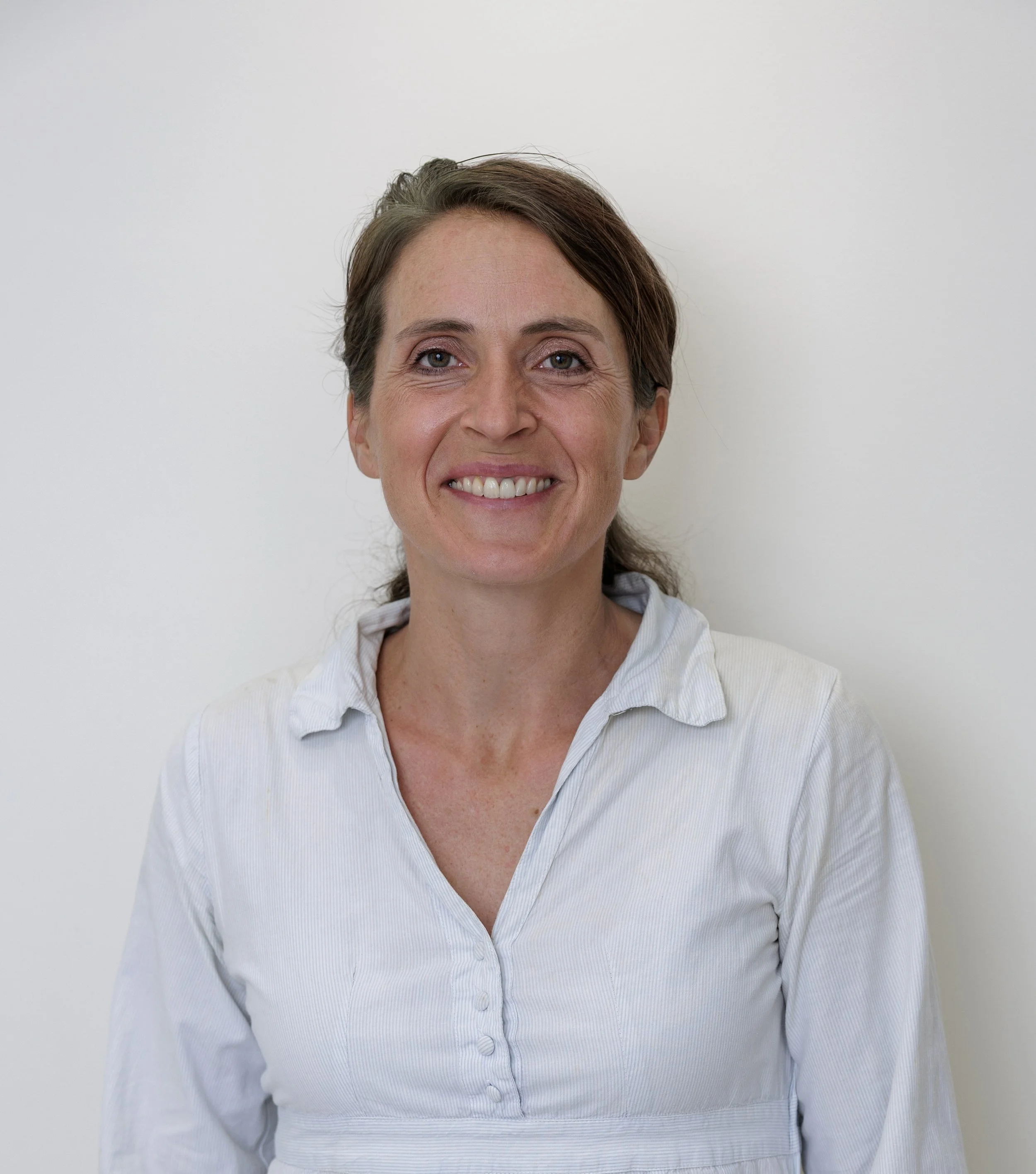
About me
-
2024- present
Private Practice, Family Medical Centre & Antiaging Clinic, Lagos- Portugal
2021- present
Private Practice, Clinica Medin, Lagoa- Portugal
2019- present
Private Practice, Family Medical Centre & Antiaging Clinic , Quinta do Lago- Portugal
2012- present
Klinik Manhagen, Hamburg- Germany
2017- 2025
Private Practice, German Medical Centre, Carvoeiro- Portugal
2017
Specialist Exam in Orthopedic and Trauma Surgery
2015- 2017
Private Practice, Orthopädiezentrum Kurfürstendamm, Berlin- Germany
2014
Further Education in osteopathic medicine with certificate „doctor of osteopathy“
DO of the german/american organisation DAAO
2012- 2013
Department of Shoulder Surgery, Sports- Trauma- and Arthroscopic Surgery,
Roland Klinik Bremen- Germany
2010- 2012
Department of Orthopedic and Trauma Surgery, Asklepios Klinik Nord Heidberg- Germany
2009- 2010
Department of Anesthesia and Intensive Care, Sana Klinikum Sommerfeld- Germany
2009
PhD at University Hamburg- Germany
2008- 2009
Department of Arthroplasty, Sana Klinikum Sommerfeld- Germany
2002- 2008
Medical school at University of Hamburg and Tauranga, New Zealand
-
Specialist in Orthopedics and Trauma Surgery
Doctor of osteopathy (DAAO)
Certified FDM- Therapist (fascial distorsion model of Typaldos)
Manual Medicine MWE
Foot Surgery GFFC
Emergency Medicine
ATLS Instructor (advanced trauma life support)
Certified 800hrs yoga teacher
-
German
English
Portuguese


Contact me directly

Specialty
Treatment of Osteoarthritis
Hyaloronic Acid vs
Platelet Rich Plasma vs
Autologous Conditioned Serum
Diagnosing joint wear is one of the most common diagnoses in orthopedic practice. The knee joint, hip joint, and hands are particularly susceptible to wear, but all other joints can also restrict the patient's quality of life due to pain and limited movement.
Typical symptoms of joint wear include pain upon starting and loading, especially in the morning and after prolonged activity, often accompanied by stiffness. The pain and stiffness usually improve after some initial movement in the morning, but can reoccur with prolonged activity. In advanced stages, patients may also experience pain at rest and during the night.
The typical restriction of movement results from the reduction of cartilage in the joint and changes in the joint capsule, tendons, and muscles surrounding the joint.
Diagnosing joint wear involves careful patient questioning, physical examination, and, if necessary, an X-ray.
There are various treatment approaches available. Maintaining a normal body weight and engaging in non-excessive or aligned movements often results in pain reduction.
Sports that unload the affected joint, such as cycling or swimming, are recommended. Walking on soft surfaces like forest terrain or grass with the use of forearm crutches or walking sticks can also provide relief.
Physical measures such as ice/cold applications for acute pain or swelling and heat applications on the surrounding muscles are perceived as pleasant and relieving.
Physical therapy or f.e. acupuncture are important accompanying therapies. Acute pain and severely inflamed tissue during an arthritis flare-up can be temporarily treated with non-steroidal anti-inflammatory drugs, if there are no contraindications (such as certain heart or gastrointestinal diseases or blood clotting disorders).
If these measures do not provide sufficient relief, injection treatments into the affected joint can be considered.
Three different therapies are distinguished in general.
Injection treatment with "platelet rich plasma" has shown good results for mild to moderate joint wear. In this case, blood taken from the arm vein, is treated through centrifugation and prevention of coagulation to extract the plasma from the blood. The plasma, containing platelets and anti-inflammatory and growth-promoting components, is then injected into the joint under sterile conditions, where it can act on the cartilage and often inflamed synovial membrane. This therapy also works very well for overloaded or chronically inflamed tendons, such as tennis elbow, Achilles tendon irritation, and muscle injuries.
In cases of moderate or severe changes in joint cartilage, the lubrication, shock absorption, and nutrition functions of the synovial fluid can be improved with hyaluronic acid injections. Typically, injections are given once and can last for 6-12 months.
In severe cases of osteoarthritis where the inflammation of the joint is the main problem and/or the hyaluronic acid infiltration has not been successful there is the option to inject ‘ autologous conditioned serum’ where the patients blood is incubated which increases the anti-inflammatory purposes of the later extracted serum.
Surgery with the implantation of an artificial joint should only be considered in cases of so-called therapy resistance, when the treatments have not been effective and when the patient expresses a strong desire for it. Ultimately, the decision maker is always the patient, who weighs the benefits for their quality of life against the risks of the outcome of an operation.
Specialty
Orthokine® Therapy – autologous conditioned serum - a treatment for osteoarthritis and chronic inflammation of the musculoskeletal system
Orthokine® therapy is a modern biological treatment that uses the natural anti-inflammatory properties of the patient’s own blood. The aim is to reduce inflammatory processes, relieve pain and thereby improve the mobility and function of the joints and spine.
An important advantage is that the substances used for treatment are obtained from the patient’s own blood. No cortisone is used as part of the treatment.
How does Orthokine® therapy work?
First, a small amount of blood is collected into a special Orthokine® syringe containing small glass beads that come into contact with the blood.
The filled syringe is then incubated for several hours at 37 °C. During this time, blood cells are stimulated to produce increased levels of the body’s own anti-inflammatory substances. One of the most important is interleukin-1 receptor antagonist (IL-1Ra). This protein acts as a natural antagonist of interleukin-1 and can inhibit an important inflammatory pathway involved, among other conditions, in osteoarthritis.
After incubation, the blood is centrifuged to separate the cellular components from the serum. The resulting Autologous Conditioned Serum (ACS) contains increased concentrations of various naturally occurring proteins and is then injected directly into the affected area.
Orthokine® may be used for a range of painful inflammatory and degenerative conditions of the musculoskeletal system. One of its main areas of application is advanced osteoarthritis of both large and small joints, as well as degenerative and osteoarthritis-related conditions of the spine.
In selected cases, the treatment may also be considered for chronic bursitis and inflammatory irritation of nerve structures, for example in association with a herniated disc.
The appropriate treatment and injection technique depend on the exact cause and location of the symptoms.
Orthokine® or PRP – what is the difference?
Both Orthokine® and PRP (Platelet-Rich Plasma) are prepared from the patient’s own blood. However, the preparation process and therapeutic focus are different.
PRP primarily concentrates platelets and the growth factors they contain. These can support tissue healing and regeneration. PRP is therefore frequently used for tendon, muscle and ligament injuries, as well as earlier stages of osteoarthritis.
Orthokine®, in contrast, focuses more strongly on modulating inflammatory processes. Incubating the blood produces a serum containing increased concentrations of the body’s own anti-inflammatory proteins.
For this reason, Orthokine® may be a particularly interesting biological treatment option for advanced or chronically inflamed osteoarthritis, as well as for degenerative conditions of the spine.


Specialty
Golfers and Tennis Elbow
The frequent medical complaint of a tennis elbow with pain on the outer side of the elbow (called epicondylitis humeri radialis) and the slightly less common problem of a golfer’s elbow with pain on the inner part of the elbow (epicondylitis humeri ulnaris) are caused by painful tissue changes in the tendons of the elbow through repetitive movement of the wrist- and hand extensors and flexors.
Often this complaint is caused by playing tennis and golf but this phenomenon is also found in people working in maintenance, long hours on the computer keyboard, in cleaning jobs, athletes in ball sports, but it is also more generally caused by intense monotonous straining of the corresponding muscles of the forearm. An uncommon exposure (f.e. a house renovation) or a rapid increase of a particular strain (f.e. starting training after a break) are the cause of the complaints. This strain might lead to inflammation of the sinewy area at the transition of bones to muscle structure at the elbow.
Not only the shortening and the hypertension of the muscles of the forearm, also a missing stabilization and unfavorable coordination of the shoulder girdle or changes in the cervical spine caused by degeneration or blockages lead to the longer lasting symptoms of epicondylitis.
Pain caused by inflammatory processes and tiny injuries, so called micro-injuries, initially start while straining the arm and later on will also occur in resting mode. Typically the pain radiates along the muscular tissue to the forearm.
A diagnosis can already be made with a thorough examination. In the examination and in the subsequent treatment it is important to include the shoulder girdle, the cervical spine and the nerve function.
The treatment should be holistic. Measures to ease the pain like local heat or cold, ointment with anti-inflammatory creams, f.e. with comfrey extract or nightly curd compresses are beneficial. Plant-based anti-inflammatory preparations to take internally, like bromelain or curcurmin and in persistent cases also the intake of traditional anti-inflammatory painkillers like Ibuprofen is commonly advised.
Accompanied by manual treatment of the tendon insertions and treatment of tensed muscles by a physiotherapist, preferably in combination with physical methods like ultrasound therapy.
Special exercises for stretching and relaxation are important, also in form of daily self practice (concentric muscle training). The increasingly popular fascia roll is able to support the relaxation of tensed muscles and will help to continue the work of the therapist.
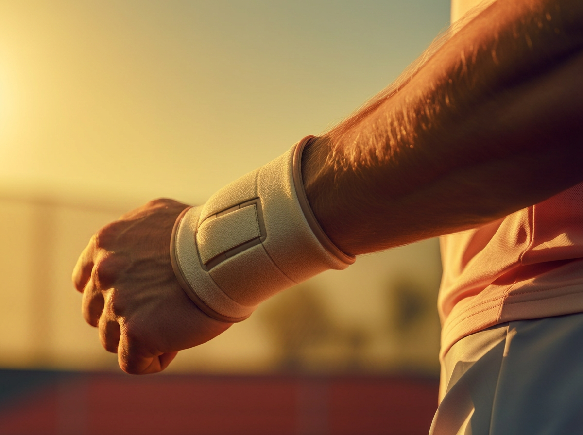
Also, the so called epicondylitis-brace, worn close to the elbow on the forearm, diverts the pull of the muscles from the origin at the elbow to less strained areas and can provide relief.
Well applied kinesiotapes bring about an increased blood circulation of the affected area and increase a relaxation of the strained muscles underneath through permanent relocation of connective tissue.
Athletes should always have their techniques inspected by an experienced coach in order to correct postural deficits.
Osteopathy with local and holistic treatment is very helpful to shorten the duration of symptoms which often last several months.
A set of specific exercises to practice at home are often passed on to the patient.
Shots of cortisone and operations meanwhile are normally not recommended any more.
For athletes a slow progression of strain of any kind (so called pacing) and continuous stretching and relaxation of muscles proved very effective to prevent recurring injuries.
In long histories of complaints injections with platelet rich plasma extracted from the patients own blood show excellent results to speed up the healing.
Specialty
Dry Needling – Targeted Treatment for Muscular Pain
Neck, shoulder and back pain are often caused or aggravated by muscular tension and myofascial trigger points. Long hours of sitting, computer work, poor posture, repetitive movements, sports and stress can all contribute to these problems.
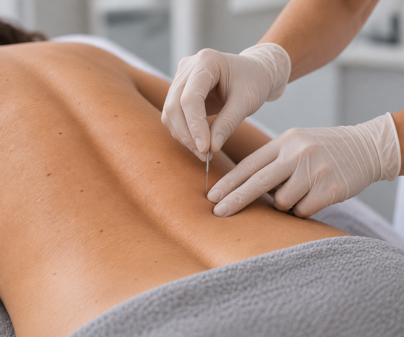
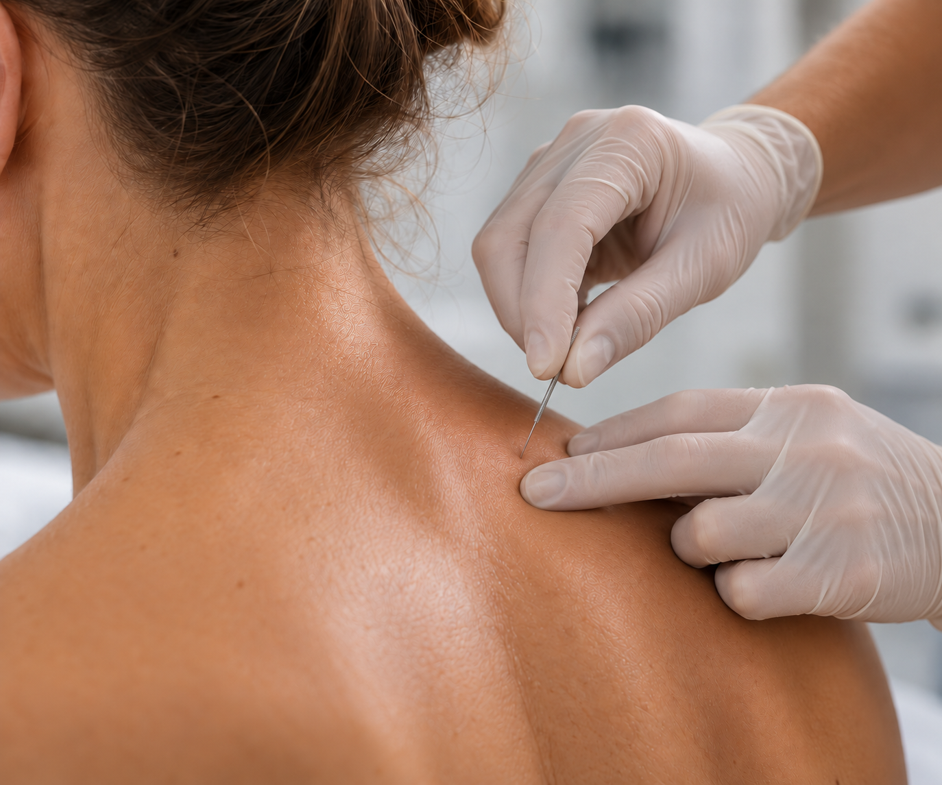
Dry Needling is a targeted treatment of myofascial trigger points using very fine, sterile needles. No medication is injected. The needle is placed directly into the affected muscle and trigger point to help reduce tension, relieve pain and restore normal muscle function.
Trigger points are small, highly sensitive areas within tense muscle fibres. They may cause pain directly in the affected muscle, but they can also produce pain in another area. For example, trigger points in the neck and shoulder muscles can contribute to headaches or pain radiating towards the shoulder and arm. Trigger points in the gluteal muscles or piriformis may cause deep buttock pain and symptoms extending into the leg.
Dry Needling can be particularly helpful for neck and shoulder pain, tension of the shoulder girdle and muscles around the shoulder blade, lower back and gluteal pain, Piriformis Syndrome, tennis elbow and muscular forearm pain. It can also be used for tension-related headaches, sports-related muscular overload and persistent muscular tension with restricted mobility.
Dry Needling is different from acupuncture. The treatment is based on anatomy, muscle function and the identification of specific myofascial trigger points.
Muscular pain often develops because of poor posture, weakness, repetitive movements or muscular overload. Dry Needling can help relax the painful muscle, but identifying and treating the underlying cause is equally important. Treatment can therefore be combined with stretching, strengthening, physiotherapy and correction of posture and movement patterns.
The aim is to reduce pain, improve muscle function and restore mobility. A careful orthopaedic examination helps determine whether the symptoms are primarily muscular or whether joints, discs or nerves are also involved.
Specialty
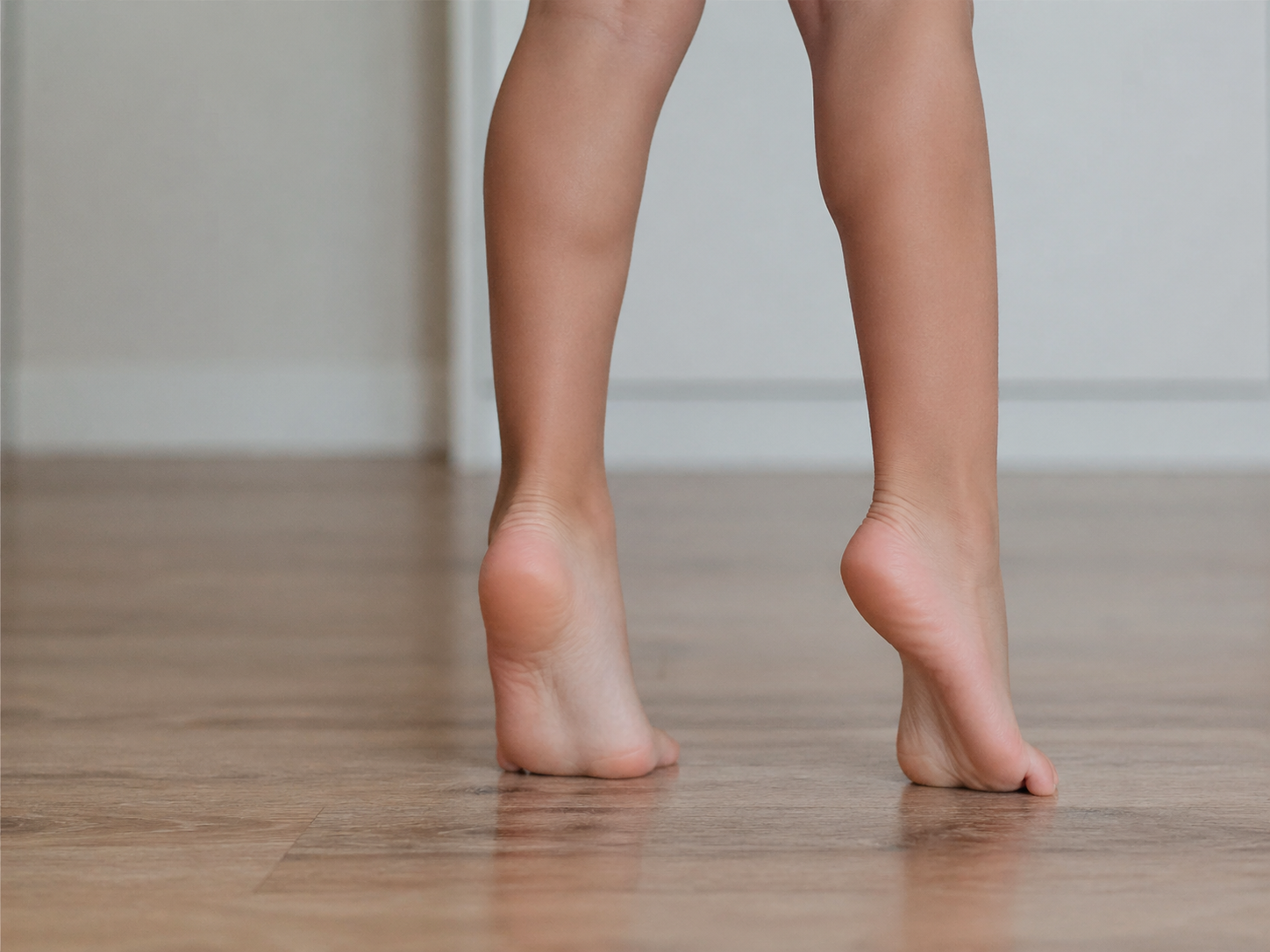
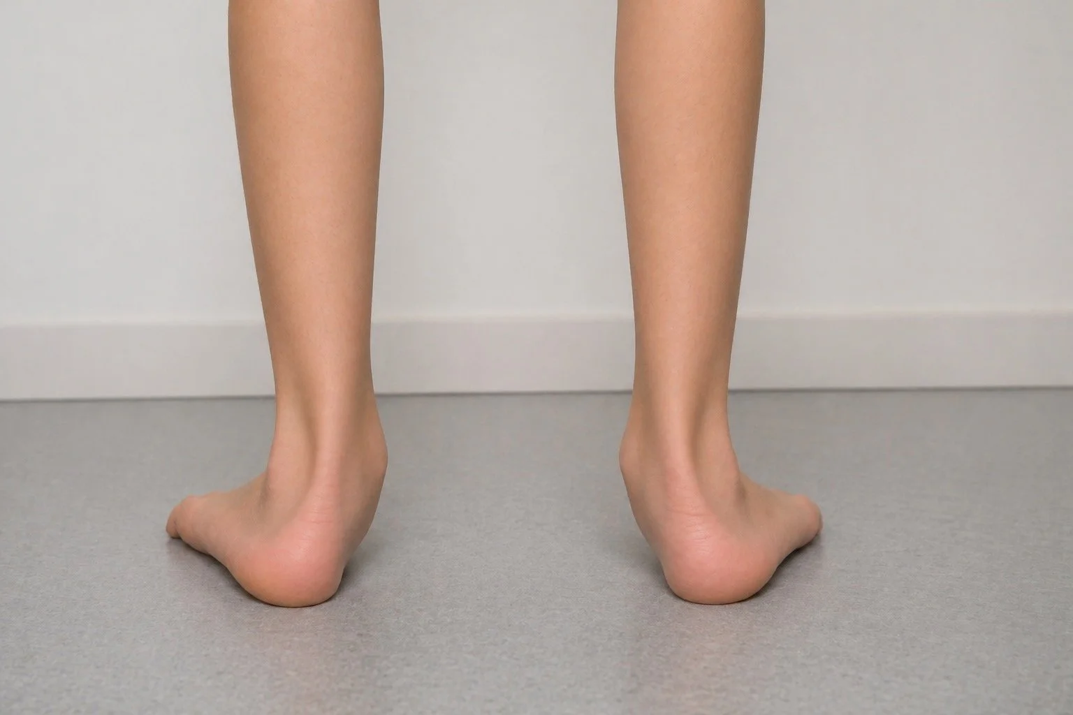
Toe Walking in Children
Many children walk on their toes at times. In early childhood, this can be part of normal motor development. However, if toe walking continues over a longer period or becomes more noticeable as the child grows, a targeted medical evaluation is recommended.
For a long time, so-called “idiopathic toe walking” was considered simply a habit. More recent scientific findings – including the work of David Pomarino – suggest that neurological, neuromuscular, or genetic factors often play an important role in affected children.
Toe walking initially describes only a noticeable walking pattern: the child mainly walks on the front of the foot without fully placing the heel on the ground. There can be many different underlying causes.
Persistent toe walking may occur in connection with:
hereditary motor and sensory neuropathies (e.g. Charcot-Marie-Tooth disease)
hereditary muscle diseases or myopathies
mild forms of cerebral movement disorders
abnormalities in muscle tone
autism-related movement patterns
sensory integration difficulties
Why Early Diagnosis Matters
Long-standing toe walking can affect overall physical development. Over time, this may lead to:
shortening of the calf muscles and Achilles tendon
reduced ankle mobility and impingement of the ankle joint in adulthood
changes in foot structure
postural differences
knee, hip, or back pain
balance and walking difficulties
The earlier underlying causes are identified, the more targeted treatment can be.
Modern assessment therefore includes not only gait observation but also neurological, orthopedic, and functional evaluation.
Therefor in children with toe walking, we perform a comprehensive clinical assessment, including:
movement patterns and gait analysis
muscle tone and strength
joint mobility
coordination and balance
neurological signs
family history
indications of neuromuscular conditions
If necessary, we perform genetic testing to identify possible causes early.
Treatment is always based on the underlying cause and the child’s individual development. The goal iis not only to change the walking pattern but to improve overall motor function.
Depending on the findings, treatment may include:
Night splints and specific insoles
specialized physiotherapy
interdisciplinary developmental assessment
minimal invasive surgery
Early and well-differentiated evaluation can help prevent functional limitations and support healthy motor development in the long term.